What osteochondritis dissecans actually is
Last reviewed November 17, 2026. OwieClear editorial team. Waiting for clinician review. Not medical advice. Talk with your doctor about your own care.
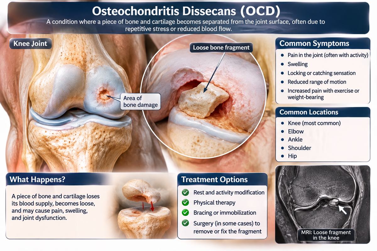
Osteochondritis dissecans is a small, focal problem with large consequences. A patch of bone just beneath the joint surface, usually a centimetre or two across, loses its blood supply and begins to separate, taking the cartilage above it with it.
If it heals, the joint is normal. If the fragment detaches, the joint has a hole in its surface and a loose piece rattling around in it, and the risk of early arthritis rises sharply.
Related pages: Osteochondritis dissecans (loose piece of bone in a joint)
The bottom line
- It affects the bone under the cartilage, not the cartilage itself at first.
- The knee is by far the most common site, then the elbow and the ankle.
- It mainly affects active children and teenagers.
- Lesions in growing children heal much better than lesions in adults.
- A detached fragment can cause locking and leads to early osteoarthritis if left.
- What actually triggers the loss of blood supply.
- Why some lesions heal and others do not.
- How much of the risk is genetic.
The story, step by step
What the evidence labels mean
What each word means
What happens
A misleading name, used anyway.
What the research shows
- Osteochondritis means inflammation of bone and cartilage, and dissecans means separating. Neither is quite right.
- There is usually no significant inflammation, and the primary problem is in the bone beneath the cartilage rather than the cartilage itself.
- The term was coined by Franz Konig in 1887, when the mechanism was unknown, and it stuck.
- Clinicians often shorten it to OCD, which causes confusion with obsessive-compulsive disorder, so the full term or OCD lesion is usually written out.
- A detached fragment floating in the joint is traditionally called a joint mouse, because it moves around and turns up in different places.
- The useful way to think of it is a small area of bone under the joint surface that has lost its blood supply and may take the cartilage above it with it.
Why it matters
The name describes neither the location nor the mechanism correctly, which is worth knowing before trying to work out the condition from the words.
What's still uncertain
- Why the original description persisted.
Read more
- NIAMS: Osteochondritis dissecansGovernment
- MedlinePlus: Osteochondritis dissecansGovernment
- American Academy of Orthopaedic Surgeons: OrthoInfoNonprofit
Next: Where it happens
Knee, elbow, ankle
What happens
Specific spots within specific joints.
What the research shows
- The knee accounts for the large majority of cases, and the classic site is the lateral side of the medial femoral condyle, the inner knuckle of the thigh bone.
- The elbow is next, typically the capitellum, the rounded part of the outer upper arm bone where it meets the radius. This is strongly associated with throwing and with gymnastics.
- The ankle, usually the dome of the talus, is third, and is often associated with previous ankle sprains or fractures.
- Less commonly the hip, shoulder, wrist and the kneecap are affected.
- Lesions are usually single, though a minority of people have more than one, sometimes in both knees or both elbows.
- Both sides are affected in perhaps a fifth of knee cases, which is one reason the other side is often imaged when one is found.
Why it matters
Knowing the usual sites explains why a thrower's elbow and a footballer's knee are the two classic presentations.
What's still uncertain
- Why these particular spots are vulnerable.
Read more
- NIAMS: Osteochondritis dissecansGovernment
- MedlinePlus: Knee injuries and disordersGovernment
- MedlinePlus: Elbow injuries and disordersGovernment
Next: Who gets it
Active young people
What happens
The age pattern is central.
What the research shows
- It most commonly appears between about 10 and 20 years of age, in physically active young people.
- Boys are affected more often than girls, although the gap has narrowed as girls' sports participation has risen.
- It is strongly associated with repetitive, high-impact or high-torque activity: football, basketball, gymnastics, baseball pitching, tennis, dance.
- Single-sport specialisation and year-round training raise the risk, because they remove the recovery periods that allow bone to remodel.
- It is uncommon but not rare. Estimates vary, in the region of 15 to 30 cases per 100,000 people.
- It can be found incidentally on imaging done for another reason, with no symptoms at all.
Why it matters
The concentration in young single-sport athletes is why this condition keeps coming up in discussions of youth sports workload.
What's still uncertain
- Whether rates are rising with early specialisation.
- Why boys are affected more often.
Read more
- NIAMS: Osteochondritis dissecansGovernment
- NIAMS: Sports injuriesGovernment
- MedlinePlus: Sports fitnessGovernment
Next: What it feels like
Vague, then mechanical
What happens
The symptoms depend on stability.
What the research shows
- Early on, the usual complaint is a poorly localised ache in the joint that is worse with activity and better with rest. It is easy to dismiss as growing pains or a strain.
- There may be mild swelling after activity, and tenderness over a specific spot on the joint line.
- A limp after sport, or an inability to fully straighten or bend the joint, can appear.
- If the fragment loosens, mechanical symptoms begin: catching, clicking, a feeling of something moving inside the joint, the joint giving way, or frank locking where it jams and will not move.
- Locking, catching and giving way are the symptoms that most strongly suggest an unstable or detached fragment, and they change the treatment.
- Symptoms can be intermittent, settling for weeks and returning, which delays diagnosis.
Why it matters
Persistent activity-related joint pain in a young athlete, lasting weeks, is the presentation that should prompt imaging rather than more rest.
What's still uncertain
- How to shorten time to diagnosis.
- Why symptoms correlate poorly with lesion size.
Read more
- MedlinePlus: Osteochondritis dissecansGovernment
- NIAMS: Osteochondritis dissecansGovernment
- American Academy of Orthopaedic Surgeons: OrthoInfoNonprofit
Next: Why it matters
The joint surface
What happens
A small defect in a load-bearing surface.
What the research shows
- If the lesion heals, which happens often in growing children, the joint surface is restored and the long-term outlook is good.
- If the fragment detaches, it leaves a crater in the joint surface. Cartilage does not regenerate, so that crater is permanent unless repaired.
- The loose fragment itself can damage the opposing joint surface as it moves around, and can jam the joint.
- A defect in a weight-bearing surface concentrates load on its edges, which accelerates wear in the rest of the joint.
- Osteochondritis dissecans is a recognised cause of osteoarthritis appearing decades earlier than usual, sometimes in the thirties or forties.
- This is why a stable lesion in a child is treated seriously despite modest symptoms: the aim is to get it to heal before it separates.
Why it matters
The whole logic of treatment is preventing a small reversible problem from becoming a permanent hole in a joint surface.
What's still uncertain
- How often early osteoarthritis follows.
- Whether cartilage repair techniques prevent it.